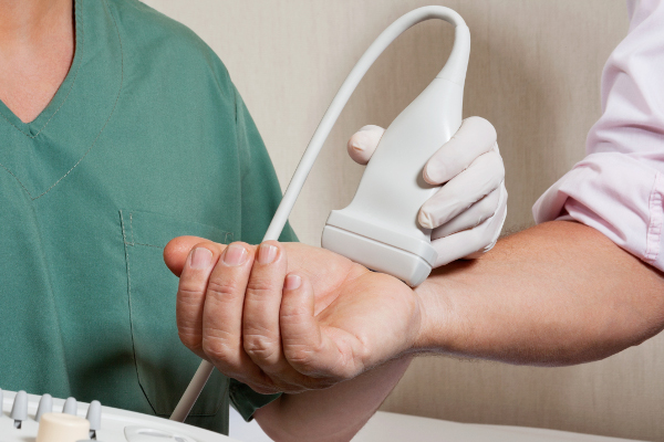
What is Musculoskeletal Ultrasound?
Musculoskeletal ultrasound is a state of the art and powerful imaging tool used for the diagnosis of various types of soft tissue injuries. It uses sound waves to create images of areas of potential injury and is radiation free and safe to use in children, adults and pregnant women. The high resolution images can assess tendon, ligaments, nerves, muscles, bursa and other soft tissues. Live action imaging can also be performed where tendons and joints can be actively moving and evaluated during ultrasound, helping better examine areas of injury.
What are the advantages of musculoskeletal ultrasound?
- Safe - Ultrasound imaging is pain free, radiation free, and non-invasive.
- Fast – It can be done in the medical office at the time of initial evaluation without a separate appointment.
- Accurate – In experienced hands, it can greatly improve accuracy of diagnosis and help guide treatments such as injections. It is performed by the physician who knows the patient history and exam, not by a technician.
- Dynamic – Unlike an MRI, live action images can be obtained showing the function and movement of the potentially injured area.
- Affordable – Ultrasound exams cost much less than MRI and CT scans.
Ultrasound guided injections
Musculoskeletal ultrasound can be used to accurately and safely guide the needle during cortisone, hyluronic acid (Synvisc, Supartz, etc), and PRP (Platelet Rich Plasma) injections. Critical structures such as nerves and blood vessels can be safely avoided and the needle can be quickly guided to its intended target for treatment. Scientific studies have clearly shown ultrasound guided injections to be superior in accuracy and comfort (less painful) than blind injections.
What are the limitations of musculoskeletal ultrasound?
Because sound wave cannot penetrate bone, any structure that is covered by bone and cannot be uncovered by moving the bone out of the way by body and limb positioning cannot be assessed by ultrasound. Examples of such structures would include the knee anterior cruciate ligament, shoulder labrum (only the back part can be seen), and spinal cord and disc issues.
Why choose us?
Dr. Ooi is one of the most experienced physician musculoskeletal ultrasonographers in the Sacramento region, being one of the first to start implementing it in his practice. He has attended many courses taught by pioneers of musculoskeletal ultrasonography and continues to stay up to date on the latest developments and techniques. He has performed diagnostic musculoskeletal ultrasound on thousands of patients and used ultrasound to guide thousands of injections.